
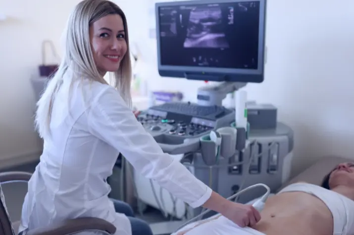
Healthcare practitioners utilize abdominal ultrasonography as a strong diagnostic technique to evaluate the organs and tissues within the abdomen.
Using sound waves to provide real-time pictures of the abdominal organs, this non-invasive imaging method offers important insights into a range of medical disorders.
You may also like:
- Is an Empty Stomach Necessary for Ultrasound?
- Why Your Practice Needs Portable Ultrasound Machines
- Medical Ultrasonography: How Does It Work and Why You Might Need It?
5 Health Conditions Abdominal Ultrasounds Can Detect
Ultrasound of the abdomen can discover kidney stones and detect abnormalities in the liver, among other medical conditions. They are an essential diagnostic tool for a broad variety of situations.
1. Gallstones and Gallbladder Disease
When evaluating the health of the gallbladder and looking for gallstones, abdominal ultrasonography is a very useful tool. Small, hardened deposits called gallstones can develop in the gallbladder and can cause symptoms including nausea, jaundice, and abdominal discomfort.
Healthcare professionals can see the gallbladder and determine if gallstones are present, as well as any inflammation or anomalies, via an abdomen ultrasound. Abdominal ultrasonography can evaluate the gallbladder’s general anatomy and function in addition to identifying gallstones.
Utilizing ultrasonic imaging, diseases like cholecystitis (gallbladder inflammation) and biliary blockage can be identified.
2. Liver Abnormalities
When it comes to assessing liver health and identifying different types of liver problems, abdominal ultrasonography is a useful diagnostic technique.
Ultrasound imaging can provide precise information about the size, shape, and texture of the liver, letting medical professionals evaluate its general function and spot any areas of concern—whether it’s fatty liver disease, liver cysts, or liver cancers.
Furthermore, fluid-filled sacs called liver cysts, which form inside the liver tissue can be seen by abdominal ultrasonography. While the majority of liver cysts are benign and asymptomatic, some have the potential to enlarge over time or manifest symptoms like discomfort or pain in the abdomen.
Healthcare professionals can use ultrasound imaging to precisely determine the size, location, and quantity of liver cysts.
3. Kidney Stones and Renal Disorders
Renal calculi, also known as kidney stones, are hard deposits that grow within the kidneys and can be very painful to pass via the urinary canal. An ultrasound of the abdomen can be used to identify kidney stones and evaluate renal function.
Ultrasound imaging can detect the existence, size, and location of kidney stones as well as any related issues such as hydronephrosis (fluid accumulation in the kidneys) by seeing the kidneys and urinary system.
Abdominal ultrasonography is a useful diagnostic tool for kidney stones as well as other renal illnesses such as renal cysts and polycystic kidney disease (PKD). A hereditary condition called polycystic kidney disease (PKD) is marked by the growth of many fluid-filled kidney cysts, which, over time, can cause kidney enlargement and reduced renal function.
Ultrasonography of the abdomen can show the kidneys and identify cysts, which helps in early PKD diagnosis and treatment.
4. Pancreatic Disorders

The diagnosis and evaluation of several pancreatic conditions, such as pancreatitis, pancreatic cysts, and pancreatic malignancies, depend heavily on abdominal ultrasonography.
Deviations in the shape or function of the pancreas, an essential organ that produces insulin and digestive enzymes, can have serious health consequences.
Additionally, pancreatic tumors, such as pancreatic adenocarcinoma, the most prevalent kind of pancreatic cancer, can be seen using ultrasound of abdominal. It is essential to discover pancreatic cancers early to start therapy on time and enhance patient outcomes.
Ultrasound imaging aids medical professionals in determining the size, location, and involvement of adjacent blood vessels of tumors, which informs treatment planning and prognosis. The imaging technology visualizes the pancreas and surrounding tissues.
5. Abdominal Aortic Aneurysms
The body’s major artery, the aorta, can develop abnormal bulges or enlargements called abdominal aortic aneurysms (AAAs), which can be seen using an ultrasound of the abdomen.
Although AAAs often show no symptoms, they can have major side effects, including rupture, which is an emergency that might be fatal. Consequently, to avoid problems and guarantee prompt action, early identification and monitoring of AAAs are crucial.
Healthcare professionals can see the abdominal aorta and see any enlargement or dilatation that might be an AAA during medical imaging like an abdominal ultrasound.
Ultrasound imaging assists in identifying the aorta’s general shape and diameter, which impacts the likelihood of a rupture and informs treatment choices like surgical repair or monitoring.
Conclusion
Versatile imaging modalities, such as ultrasound of the abdomen, are essential for detecting and tracking a wide range of health disorders affecting the organs and tissues of the abdomen.
Ultrasound imaging offers important insights that inform treatment choices and enhance patient care, from diagnosing abdominal aortic aneurysms and gallstones to evaluating kidney function. See a medical expert for further assessment and suitable treatment if you exhibit symptoms or have risk factors for any of these illnesses.
About The Author:
Stacey Smith is a freelance health writer. She is passionate about writing about women’s health, dental health, diabetes, endocrinology, and nutrition and provides in-depth features on the latest health news for medical clinics and health magazines.


