Neurological conditions are now among the most common health problems these days. These affect the spine, nerves, and brain and include memory disorders, epilepsy, migraines, brain tumors and aneurysms, peripheral neuropathy, and ALS, among others.
In the past, the diagnosis of these conditions was primarily based on a patient’s symptoms. That said, most people ordinarily assume that doctors have made a misdiagnosis when they are told they have a neurological condition. Neurology centers in St George and other metropolitan areas now have modern medical equipment to provide a proper diagnosis.
Neurology is, however, not a guesswork treatment. There are now various diagnostic tests used to determine the condition you are suffering from based on the symptoms you experience. Among the standard tests are the imaging studies.
The following are some of the imaging tests used to determine neurological conditions:
PET scanning
Positron emission tomography (PET) scanning measures the sugar level in your brain. It will illustrate the point where neural misfiring happens. This test is based on the fact that your active neurons will use glucose as the fuel for their operation. A tracer substance will be injected into your bloodstream and create visible spots that will be picked by detectors. The detectors will then generate a video of your brain when you are active. This imaging test can be used to diagnose tumors and seizure and memory disorders. PET scans are, however, not routinely used since they are expensive and invasive.
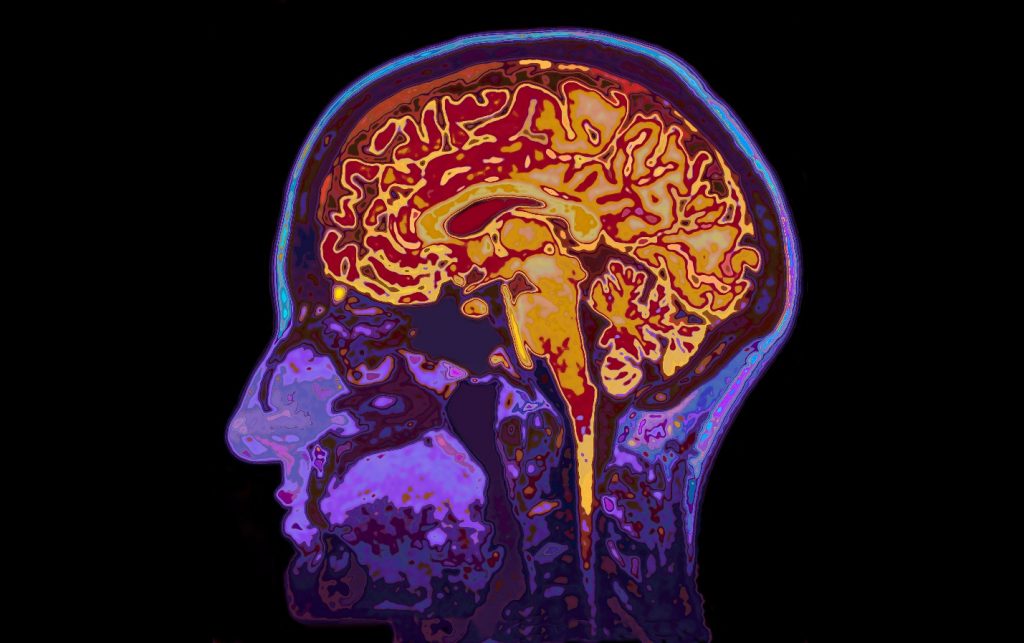
Magnetic Resonance Imaging (MRI)
This uses a strong magnetic field for the alignment of spinning atomic nuclei within your body’s tissues. Through the process of alignment, a radio frequency will be generated. This will generate an image of your brain’s structure. MRIs are noninvasive, have minimal health risks, and can even be used in utero and on infants. They are used to diagnose tumors, torn ligaments, inflammations, infections, and spinal cord and brain abnormalities.
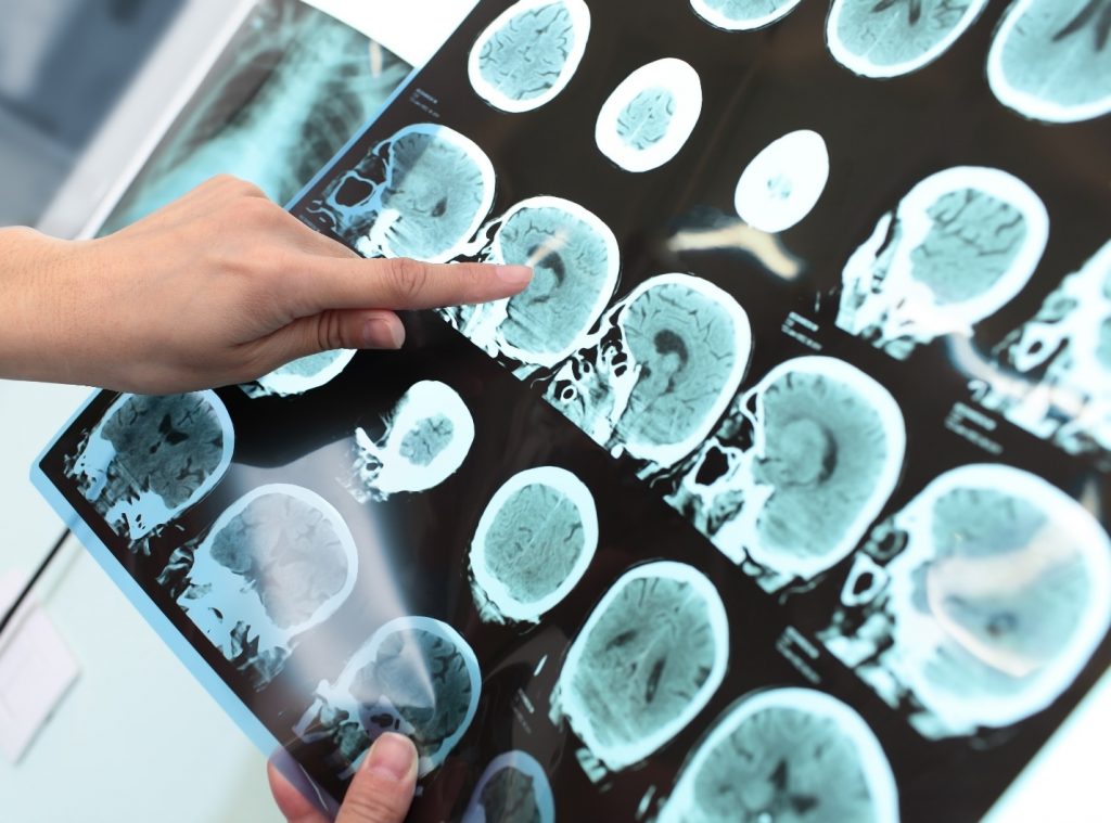
CT scan
Computerized tomography (CT) scanning uses X-rays for the production of multiple cross-section images of your brain. This test is used for the identification of broken bones, blood clots, and bleeds that might affect the functioning of your brain and other parts of your nervous system. CT scans are painless, fast, simple and have minimal radiation exposure.
Carotid Ultrasound
Carotid Ultrasound is used to determine blood clots and plaques that might impede the blood flow to the carotid artery. A gel is applied on the skin, and then a transducer passes over it to generate an image on an ultrasound screen. The test is painless and noninvasive and it takes 15-30 minutes. A carotid ultrasound can be used to pick tumors, blood clots, and any obstructions in the blood flow to your brain.
With the above technologies, a doctor will have a clear picture of your condition. He/she can then tailor your treatment to address the problem rather than give you multiple medications hoping one will work. You cannot ignore headaches, constant fatigue, muscle weakness, and reduced cognitive abilities since these might be symptoms of a neurological disorder. Get the above imaging tests to have a definite diagnosis of your symptoms and learn the right treatment.
About The Author:
Anne Kamwila is a freelance content writer and a digital marketer. She is passionate to write about health, technology, and business-related guides, news, and books.


